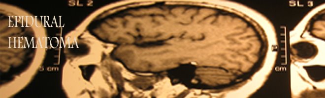
Epidural Hematoma usually results from a brief linear contact force to the calvaria that causes separation of the periosteal dura from bone and disruption of interposed vessels due to shearing stress. Epidural hematoma When there is a direct blow to the head, the bruising of the brain and the damage to the internal tissue and blood vessels is due to a mechanism called coup-countercoup. An Epidural Hematoma occurs when a blood clot forms underneath the skull, but on top of the dura, the tough covering that surrounds the brain.
Skull fractures occur in 85-95% of adult cases. Arterial or venous structures may be compromised, causing rapid expansion of the hematoma; Extension of the hematoma usually is limited by suture lines owing to the tight attachment of the dura at these locations. Recent analyses have revealed that epidural hematoma may actually traverse suture lines in a minority of cases.
The temporoparietal region and the middle meningeal artery are involved most commonly (66%), although the anterior ethmoidal artery may be involved in frontal injuries, the transverse or sigmoid sinus in occipital injuries, and the superior sagittal sinus in trauma to the vertex. Bilateral epidural hematomas account for 2-10% of all acute epidural hematomas in adults but are exceedingly rare in children. Posterior fossa epidural hematomas represent 5% of all cases of epidural hematoma.
Patients with this type of condition frequently have bruises around their eyes and a bruise behind their ear. They may also have clear fluid draining from their nose or ears due to a tear in part of the covering of the brain. These patients usually require close observation in the hospital.
Spinal epidural hematoma may be spontaneous or may follow minor trauma, such as lumbar puncture or epidural anesthesia. Spontaneous spinal epidural hematoma may be associated with anticoagulation, thrombolysis, blood dyscrasias, coagulopathies, thrombocytopenia, neoplasms, or vascular malformations. The peridural venous plexus usually is involved, though arterial sources of hemorrhage also occur. The dorsal aspect of the thoracic or lumbar region is involved most commonly, with expansion limited to a few vertebral levels.
The full extent of the problem may not be completely understood immediately after the injury, but may be revealed with a comprehensive medical evaluation and diagnostic testing. The diagnosis of a head injury is made with a physical examination and diagnostic tests. During the examination, the physician obtains a complete medical history of the patient and family and asks how the injury occurred. Trauma to the head can cause neurological problems and may require further medical follow up.

Epidural Hematoma

epidural hematoma.

No comments:
Post a Comment